Diagram Of Epidermis Of Onion Leaf
Onion epidermal cell labeled diagram Onion cells cell micrograph epidermis bulb stock alamy polars containing crystals Onion leaf epidermis with stomata pores
Onion Epidermal Cell Diagram
Cells artificial epidermis epidermal Onion cells epidermal cell red epidermis peel wikipedia size Onion epidermal cell diagram
Onion epidermis slide biology practical prepare 9th
Onion cells microscope hi-res stock photography and imagesOnion cell epidermal diagram labeled cells microscope under drawing skin epidermis lab bulb mag membrane observation vacuole nucleus leaves preparation Epidermis onion bulbo escala cebola epiderme cebolla lampadina epidermide cipolla scala pilhas archivo- gold-plated onion cells make artificial muscles.
Onion epidermal cellsOnion epidermal drawings epidermis labeled biology chromosomes chromosome dna observation Cells epidermal epidermis micOnion under skin stained epidermis mesophyll epithelium pee ling fig showing bulb mag microscopy.

Biology practical| 9th biology practical
Stomata leaf epidermis onion poresOnion 100x epidermis Peel stained epidermal microscope observation labelledOnion cells stock photos & onion cells stock images.
Microscope epidermal cell membrane vacuole nucleus stain iodine staining molecular pngkit organelle investigating celluloseThe inner epidermis of the onion bulb’s cataphylls (the onion skin). Onion cells light micrograph photomicrograph microscope high seen through cell resolution epidermis bulb organelles stock alamy wall scaleMicroscope epidermis furian hermes 26th.

Onion epidermis with large cells under microscope photograph by peter
Onion epidermis 100x: general biology lab: loyola university chicagoOnion epidermal cell Onion bulb structure cepa allium foods red bulbs schematic figureOnion cells stock photos & onion cells stock images.
Onion bulb scale epidermis stock image. image of leafOnion cell cells microscope micrograph microscopic alamy stock skin magnification section high allium cepa mitosis root To prepare a stained, temporary mount of onion peel and to study its cells.Epidermal onion cells under a microscope.


Onion bulb scale epidermis stock image. Image of leaf - 47620127

Onion leaf epidermis with stomata pores - Stock Image - B745/0273

Onion Epidermal Cells | www.pixshark.com - Images Galleries With A Bite!

Onion epidermal cell - Wikipedia

The inner epidermis of the onion bulb’s cataphylls (the onion skin).

Foods | Free Full-Text | Structural Changes Induced by Pulsed Electric

Onion epidermis with large cells under microscope Photograph by Peter
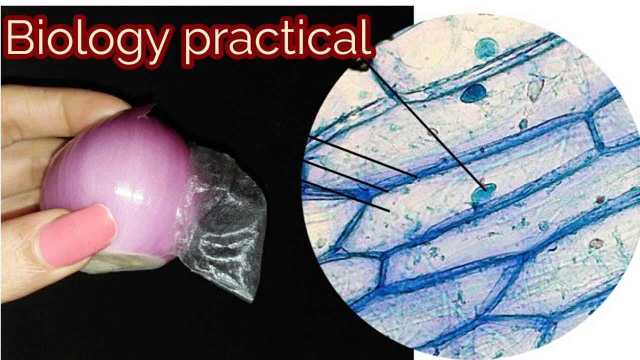
Biology practical| 9th biology practical | prepare slide of onion

Epidermal Onion Cells Under A Microscope - Different Types Of Cells